Cardiovascular Imaging
Non-invasive cardiovascular imaging has rapidly evolved over the last decades. Owing to numerous innovations, multimodality cardiovascular imaging allows nowadays reliable diagnosis, risk stratification, planning and monitoring of therapeutic interventions for all major cardiovascular diseases. Excellence in patient care is the primary aim of the interdisciplinary cardiovascular imaging center at our institution that represents the collaborative efforts of the Departments of Cardiology, the Department of Radiology and Nuclear Medicine. We are actively involved in several clinical research projects using latest technology in computed tomography, cardiac magnetic resonance and nuclear imaging. Furthermore, we have established research efforts in collaboration with the Translational Imaging Center (TIC), that provides access to a high-end 7.0 Tesla magnetic resonance at the SITEM Insel (Swiss Institute for Translational and Entrepreneurial Medicine), which is available at only very few sites in Europe, for particular research questions.
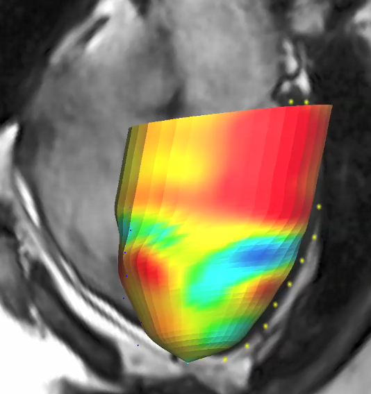
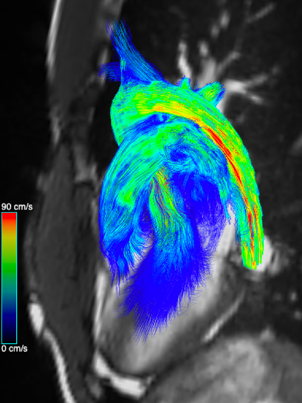
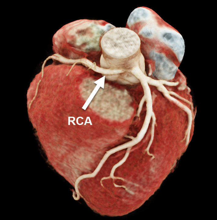
The research focus of our group investigates the application of cardiovascular computed tomography to enable optimal planning of cardiac device interventions (i.e. valvular heart disease) and to identify predictive features in non-invasive anatomical and functional imaging among patients with coronary artery disease and those with coronary artery anomalies (i.e. SNF funded project: NARCO trial). A study including patients with heart failure and preserved ejection fraction (HOPP-Bern trial) will provide insights on the development of cardiac and vascular function (strain analysis) and aortic 4D flow. In addition, we actively investigate inflammatory cardiomyopathies (i.e. cardiac sarcoidosis and peri-myocarditis) with the aim to improve early diagnosis and risk stratification using tissue characterization (depicting fibrosis with Late Gadolinium Enhancement, T1 mapping, extracellular volume fraction and edema imaging) as well as novel myocardial feature tracking strain analysis using cardiovascular magnetic resonance imaging (i.e. FlamBer registry). Similarly, we aim to improve the non-invasive diagnosis of infiltrative cardiomyopathies such as cardiac amyloidosis (i.e. B-CARE trial) to obtain a better understanding of the pathophysiology and treatment response to novel therapies. A SNF funded research project (PRE-MITRA) assesses parameters in cardiac magnetic resonance imaging to predict benefit in patients with severe mitral valve regurgitation who are undergoing edge-to-edge mitral valve repair.
Contact
Prof. Dr. Dr. med. Christoph Gräni
Director Cardiovascular Imaging
Department of Cardiology
Bern University Hospital – Inselspital
Freiburgstrasse
3010 Bern, Switzerland
031 632 45 08